Risk Factors and Treatment Fees for Implant Dentistry |
August 2011 | |
| Get the facts from implant guru Dr. Carl E. Misch as he writes about implant dentistry today. | ||
 by Carl E. Misch, DDS, MDS, PhD (hc) by Carl E. Misch, DDS, MDS, PhD (hc)Introduction Implant dentistry has become the most predictable method to replace missing teeth. However, treatment planning for implant dentistry is most often driven by the existing bone volume in the edentulous sites. This method is often problematic. In partially edentulous patients, more than 6mm of bone height is found in 40 percent of posterior maxillae and 50 percent of posterior mandibles. This percentage is further reduced to less than 20 percent of completely edentulous patients in either arch. The doctor and the patient often have an incentive to do treatment which is faster, easier and less expensive. The typical fees associated with treatment in implant dentistry are related to the number of implants and teeth replaced. Hence, a three-unit fixed partial denture supported by two implants is one-half the fee of a six-unit fixed partial denture supported by four implants. As a result, instead of bone grafts and posterior implants, distal cantilevers are often extended from anterior implants, since more vertical bone is found anterior to the maxillary sinus in the maxilla or the inferior alveolar nerve and mental foramen in the mandible.The primary cause of complications in implant dentistry is related to biomechanical factors, with too much stress applied to the implant support system. When implants are inserted into abundant bone volume and allowed to integrate for four or more months before loading, the surgical success rate is more than 98 percent. This success rate is not related to implant number, size or design. However, when the implant is occlusal loaded with the prosthesis, the failure rate might be greater than three to six times the surgical failure. For example, a meta-analysis reveals 15 percent failure rates (with several reports of more than 30 percent failure) when the implant prosthesis is occlusal loaded with implants shorter than 10mm, or when they are placed in softer bone. This failure most often occurs during the first 18 months of loading and is called early loading failure. Mechanical complications of the implant components or prosthesis outnumber surgical failures and many reports are more frequent than early loading failures. These complications include abutment screw loosening, uncemented prostheses and porcelain fracture. These complications are more often in bruxism patients, males, when opposing implant prostheses and with group function occlusion. All of the factors increase the amount of stress on the implant system (occlusal porcelain, cement, implant abutment screw and implant-bone interface). Biomechanical stress might also cause marginal crestal bone loss. Since the implant does not have a periodontal membrane as a tooth, the stress to the implant-bone interface is mostly to the crestal marginal bone. When the stress is beyond the bone physiologic limit, resorption might occur. The bone loss might increase the risk of anaerobic bacteria and peri-implantitis, or the surrounding soft tissues might shrink and result in poor cervical aesthetics. Hence, biomechanical factors can lead to early loading failure, mechanical complications and/or marginal bone loss around an implant. Stress Magnifiers Cantilevers on the prosthesis are one of the most significant stress magnifiers to the implant system. When used in the posterior regions, the greater bite force (up to five times greater than the anterior region), is further magnified and might increase the force on the implant system by three times. In order to eliminate posterior cantilevers, a bone augmentation is often indicated. Most bone augmentation procedures are not as predictable as implant integration in existing bone volumes. Bone augmentation often requires an additional surgery prior to implant placement.Additional training is required to learn bone augmentation procedures and the learning curve is longer and more difficult to become accomplished in these techniques. Complications related to bone augmentation are more common than implant surgery in existing bone volumes and might be more extensive and even debilitating to the patient. The discomfort following bone augmentation is usually more than occurs after implant surgery. An extended healing time of four to nine months might be necessary for the bone graft to mature, compared to implant healing in native bone. The costs associated with bone augmentation are often greater than the fees related to implant insertion. In addition, there are usually more implants and more teeth replaced after bone augmentation compared to situations when implants are inserted into existing volumes of the bone and teeth are cantilevered to the posterior regions. More implants and more teeth replacements further increase the cost to the patient. As a consequence of these considerations, the doctor and the patient are both motivated to use existing bone volumes for implants and restore fewer posterior teeth in the prosthesis, often with a cantilever.  Risk Factors An example of the patient and doctor having incentive to perform procedures with higher risks is when a patient has four teeth missing in a posterior maxillary quadrant (two premolars and two molars), with a pneumatized maxillary sinus cavity. There are typically two treatment options. The first is to place two implants anterior to the sinus, which supports a three-unit prosthesis (with a first molar cantilever). A second option is to perform a sinus bone graft, the insertion of three implants (in the first premolar, first molar and second molar position) and to fabricate a four-unit restoration.Fees and Risk Factors The first treatment option is one-half the fee of the second option, since it doesn't require a sinus graft, has fewer implants and less teeth replaced. The first option is also faster and easier since a bone regeneration is not required. The patient undergoes one surgery and therefore experiences less discomfort. However, the second treatment option has three to four times better chance for long-term success, since it doesn't cantilever a pontic in the molar region. Since cantilevers increase the biomechanical force to the anterior implants, there is an increased risk of an unretained prosthesis on the first premolar (because of a tensile force to the retainer and cement is 20 times weaker to tension compared to compression). This results with one implant (the second premolar) supporting three teeth and the risk of overload and failure. The first treatment option more often has more bone loss from occlusal overload related to the increased biomechanical stress as a result of the cantilever (Fig. 1). In addition, the mandibular second molar might erupt past the plane of occlusion with the first option (since it only has one molar) and each protrusive mandibular movement would result in a lateral premature contact on the maxillary prosthesis. This bone direction increases the sheer force, and might even trigger parafunction. As a result, all complications related to stress are increased. The first treatment option more often has more bone loss from occlusal overload related to the increased biomechanical stress as a result of the cantilever (Fig. 1). In addition, the mandibular second molar might erupt past the plane of occlusion with the first option (since it only has one molar) and each protrusive mandibular movement would result in a lateral premature contact on the maxillary prosthesis. This bone direction increases the sheer force, and might even trigger parafunction. As a result, all complications related to stress are increased.Biomechanical-related complications often occur within the first few years of function. As a result, the patient expects the dentist to repeat the treatment for no charge. When the first option fails, the second treatment option might be selected, often from a different dentist, which is associated with a greater cost. As a result, the patient is more likely to bring litigation against the first treatment team in order to pay for the additional costs of the second treatment option. As a consequence of an increased risk of complications in the first treatment option, the fees for this option should be more than the second treatment option. In other words, the fee for services rendered should not only be based upon the sum of the number of implants and teeth in the prosthesis, it should also include the amount of risk associated with the treatment. A more basic example of charging for risk factors is the treatment for a crown on a maxillary central incisor compared to a mandibular molar. The time and technique for an anterior preparation, impression and transitional prosthesis is greater than to restore a mandibular posterior tooth. The risk that an anterior maxillary crown has to be redone because of gingival recession, shade selection, etc. is greater than the mandibular crown. Yet, most dentists charge the same fee for both procedures. The maxillary anterior crown has more risk, therefore the fee should be greater. Full-arch Restorations When a full-arch fixed implant restoration is the treatment for a maxillary arch, the number of implants is often the same as the mandible. For example, "all in four" is a common treatment option presented to the profession in either arch, along with similar fees for either arch to the patient. Yet, the maxillary fixed restoration is supported by softer bone. The hardness of the bone is related to its strength. The mandible more often has hard (strong) bone and the maxilla most often has softer bone. In fact, the posterior maxillary bone might be five to 10 times weaker than the hard bone of the anterior mandible.The maxillary anterior arch receives a force at a 12- to 15- degree angle during occlusion and up to a 30-degree angle in excursions. A 15-degree angled force increases the force component by 25.9 percent and a 30-degree force increases the force by 50 percent. The excursive forces in a maxillary restoration come from within the arch to push outside the arch. This force direction on an arch is more detrimental than in the mandible. The mandible receives a force from outside of the arch toward the inside of the arch, which is the direction of force the arch was designed to resist. The maxillary arch usually has shorter implants than the mandible (since the vertical height of bone is less compared to the anterior mandible). The shorter implants have less surface area and higher stresses, especially in soft bone. A literature review reports a failure rate three times higher in full-arch maxillary implant fixed restorations compared to full-arch implants and a mandibular restoration. Aesthetic retreatments and speech complications are more often observed in the maxillary restoration compared to the mandible prosthesis. The air can escape between the residual ridge and prosthesis, and aesthetic requirements for the patients are primarily obtained by the maxillary restoration. In other words, the maxillary full-arch restoration should be treatment planned differently and cost more than a similar restoration in the mandible. 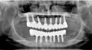 To compensate for the softer bone and higher biomechanical stress, the maxillary arch should more often have bone augmentation (to eliminate posterior cantilevers), more implants inserted and higher prosthetic fees than a mandibular arch. The fees for an implant treatment plan that has fewer implants and/or cantilevers should be greater than restorations supported by more implants and/or without cantilevers (Figs. 2-8). To compensate for the softer bone and higher biomechanical stress, the maxillary arch should more often have bone augmentation (to eliminate posterior cantilevers), more implants inserted and higher prosthetic fees than a mandibular arch. The fees for an implant treatment plan that has fewer implants and/or cantilevers should be greater than restorations supported by more implants and/or without cantilevers (Figs. 2-8).Summary The fees associated with implant surgery and prosthetic rehabilitation should be related to the risks related to the treatment. The treatment plan in implant dentistry should have a biomechanical rationale to decrease stress to the implant system. The risks in dentistry are a factor which should be included in the cost of most all procedures that are associated with greater complications. The implant and associated restoration is not a commodity in which the cost is solely related to the number of implants and prosthetic units.References 1. Goodacre CI, Bernal G, Rungcharassaeng K. Clinical complications with implants and implant prostheses. J Prosth Dent 90: 121- 132, 2003. |
||
| Author’s Bio | ||
 Carl E. Misch, DDS, MDS PhD (hc) is the professor and director of Oral Implantology at Temple Dental School in Philadelphia, Pennsylvania; and director at Misch International Implant Institute in Beverly Hills, Michigan. Carl E. Misch, DDS, MDS PhD (hc) is the professor and director of Oral Implantology at Temple Dental School in Philadelphia, Pennsylvania; and director at Misch International Implant Institute in Beverly Hills, Michigan. |
||
| Call Now San Francisco Dentist Request for appointment at: (415) 391 - 7751 450 Sutter street, Suite 1905 San Francisco, CA, 94108 http://malidds.com/ |
||

Sunday, September 4, 2011
Risk Factors and Treatment Fees for Implant Dentistry
Subscribe to:
Post Comments (Atom)
No comments:
Post a Comment